vol. 6 3/2017 Inżynier i Fizyk Medyczny
188
diagnostyka
\
diagnostics
artykuł naukowy
\
scientific paper
Literatura
1.
T. Fitzmauric, M. Naghavi, Ch.J.L. Murra:
The Global Burdern of
Cancer 2013
, JAMA Oncol., 1(4), 2015, 505-527.
2.
R. Kordek:
ONKOLOGIA, Podręcznik dla studentów i lekarzy
, Wyd.
Via Medica, Gdańsk 2007.
3.
J. Didkowska, U. Wojciechowska, W. Zatoński:
Prognozy zacho-
rowalności i umieralności na nowotwory złośliwe w Polsce do 2025
rok
,
Publikacja wydana w ramach zadania ‘Rejestracja nowo-
tworów złośliwych’ Narodowego Programu Zwalczania Chorób
Nowotworowych, Warszawa 2009.
4.
J. Meder:
Podstawy Onkologii Klinicznej
,
Centrum Medyczne
Kształcenia Podyplomowego, Warszawa 2011.
5.
.
6.
P.E. Christian, N.M. Swanston:
PET Study Guide
, SNM, Inc. United
States of America 2010.
7.
B. Małkowski:
Stan obecny i perspektywy rozwoju pozytonowej
emisyjnej tomografii w Polsce
, NOWOTWORY Journal of Onco-
logy, 57(3), 2007, 249-260.
8.
Zarządzenie Nr 67/2011/DSOZ Prezesa Narodowego Funduszu
Zdrowia z dnia 18 października 2011 r.
9.
J.W. Fletcher, B. Djulbegovic, H.P. Soares et al.:
Recommenda-
tions on the use of F-18-FDG PET in oncology
, J Nucl Med, 49,
2008, 480-508.
10.
T.A. Smith: FDG
uptake, tumour characteristics and response to
therapy: a review
, Nucl Med Commun, 19, 1998, 97-105.
11.
tynski.pdf.
12.
J. Weber, U. Haberkorn, W. Mier:
Cancer Stratification by Molecu-
lar Imaging Int. J. Mol. Sci.
, 16, 2015, 4918-4946.
13.
B. Birkenfeld, M. Listewnik:
Medycyna Nuklearna obrazowanie
molekularne
, Wydawnictwo Pomorskiego Uniwersytetu Me-
dycznego w Szczecinie, 2011.
14.
P.D. Shreve, Y. Anzai, R.L. Wahl
: Pitfalls in oncologic diagnosis
with FDG PET imaging: physiologic and benign variants
, Radio-
graphics, 19, 1999, 61-77.
15.
M.R. Habibian, J.V. Vitola, D. Delbeke, M.P. Sandler, W.H. Mar-
tin:
Nuclear Medicine Imaging: A Teaching File, 2
nd
ed.
, Lippincott
Williams & Wilkins, a Wolters Kluwer business, China 2009.
16.
M.K. Rahim, S.E. Kim, H. So, H.J. Kim, G.J. Cheon, E.S. Lee,
K.W. Kang, D.S. Lee:
Recent Trends in PET Image Interpretations
Using Volumetric and Texture-based Quantification Methods in
Nuclear Oncology
, Nucl Med Mol Imaging, 48, 2014, 1-15.
17.
M. Walentowicz-Sadłecka, P. Sadłecki, B. Małecki, P. Walento-
wicz, A. Marszałek, P. Domaracki, M. Grabiec:
Wartości SUVmax
mierzone za pomocą 18F-FDG PET/CT w guzie pierwotnym a cechy
kliniczno-patologiczne endometrialnego raka endometrium
, Gine-
kol Pol, 84, 2013, 748-753.
18.
S.H. Moon, S.H. Hyun, J.Y. Choi:
Prognostic Significance of Volu-
me-Based PET Parameters in Cancer Patients
, Korean J Radiol,
14(1), 2013.
19.
M. Vanderhoek, S.B. Perlman, R. Jeraj
: Impact of the definition
of peak standardized uptake value on quantification of treatment
response
, J Nucl Med, 53, 2012, 4-11.
20.
M. Soret, S.L. Bacharach, I. Buvat:
Partial-volume effect in PET
tumor imaging
, J Nucl Med, 48, 2007, 932-945.
21.
P. Hogg, G. Testanera:
Principles and Practice of PET/CT, Part 1,
A Technologist’s Guide
, European Association of Nuclear Medi-
cine, August 2010.
22.
A. Gallamini, C. Zwarthoed, A. Borra:
Positron Emission Tomo-
graphy (PET) in Oncology
, Cancers, 6, 2014, 1821-1889.
23.
P. Tylski, S. Stute, N. Grotus, K. Doyeux, S. Hapdey, I. Gardin,
B. Vanderlinden, I. Buvat:
Comparative assessment of methods
for estimating tumor volume and standardized uptake value in (18)
F-FDG PET
, J Nucl Med, 51, 2010, 268-276.
24.
S.M. Larson, Y. Erdi, T. Akhurst, et al.:
Tumor treatment response
based on visual and quantitative changes in global tumor glycolysis
using PET-FDG imaging: the visual response score and the change in
total lesion glycolysis
, Clin Positron Imaging, 2(3), 1999, 159-171.
25.
S. Kiyohara, S. Nagamachi, H. Wakamatsu, R. Nishii, S. Fujita,
S. Futami, et al.:
Usefulness of metabolic volume and total lesion
glycolisis for predicting therapeutic response in cancer therapy by
18 F-FDG PET/CT
, KakuIgaku, 48, 2010, 447-455.
26.
C.M. Costelloe, H.A. Macapinlac, J.E. Madewell, et al.:
18F-FDG
PET/CT as an indicator of progression-free and overall survival in
osteosarcoma
, J Nucl Med, 50(3), 2009, 340-347.
27.
H.Y. Lee, S.H. Hyun, K.S. Lee, et al.:
Volume based parameter of
(18)F-FDG PET/CT in malignant pleural mesothelioma: prediction
of therapeutic response and prognostic implications
, Ann SurgOn-
col, 17(10), 2010, 2787-2794.
28.
P. Xie, J.B. Yue, H.X. Zhao, et al.:
Prognostic value of 18F-FDG
PET-CT metabolic index for nasopharyngeal carcinoma
, J Cancer
Res ClinOncol, 136(6), 2010, 883-889.
29.
H.H. Chen, N.T. Chiu, W.H. Su, H.R. Guo, B.F. Lee:
Prognostic vale
of whole-body total lesion glycolysis at pretreatment FDG/PET-CT
in Non-Small Cell Lung Cancer
, Radiology, 264(2), 2012, 559-566.
30.
M.C. Asselin, J.P. O’Connor, R. Boellaard, N.A. Thacker, A. Jack-
son:
Quantifying heterogeneity in human tumours using MRI and
PET
, Eur J Cancer, 48, 2012, 447-455.
31.
S. Chicklore, V. Goh, M. Siddique, A. Roy, P.K. Marsden, G.J. Cook:
Quantifying tumor heterogeneity in 18 F-FDG PET/CT imaging by
texture analysis
, Eur J Nucl Med Mol Imaging, 40, 2013, 133-140.
32.
F. Tixier, C.C. Le Rest, M. Hatt, N. Albarghach, O. Pradier, J.P.
Metges, et al.:
Intratumor heterogeneity characterized by textural
features on baseline 18 F-FDG PET images predicts response in con-
comitant radiochemiotherapy in esophageal cancer
, J Nucl Med,
52, 2010, 369-378.
33.
H.P.F. van Velden, P. Cheebsumon, M. Yaqub, E.F. Smit, O.S. Ho-
ekstra, A.A. Lammertsma, R. Boellaard:
Evaluation of a cumula-
tive SUV-volume histogram method for parameterizing heteroge-
neous intratumoral FDG uptake in non-small cell lung cancer PET
studies
, Eur J Nucl Med Mol Imaging, 38, 2011, 1636-1647.
0
10
20
30
40
50
60
70
80
90
100
0
20
40
60
80
100
FDG
FLT
18F‐FDG
18F‐FLT
18F‐FDG
18F‐FLT
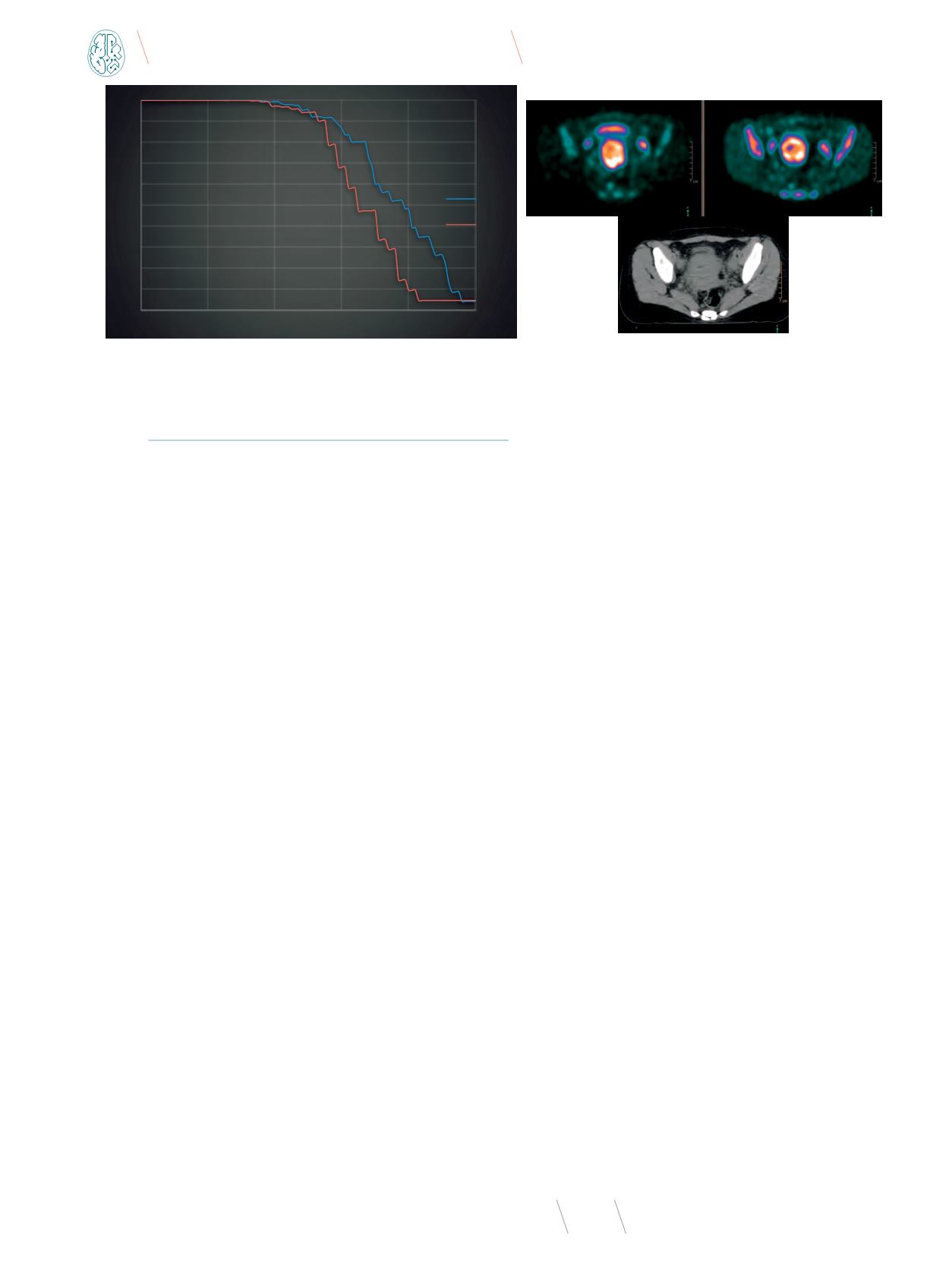
Rys. 8
Przykład rozkładu heterogenności w oparciu o AUC-CSH histogram u pacjentki z rakiem szyjki macicy przy użyciu dwóch znaczników
Źródło: Materiał własny.


